
June 5, 2019
A new discovery which may enable researchers to better comprehend the rationale behind the increased association of premature births and neurodevelopmental disorders, such as attention deficit disorders and autism spectrum disorders is that connections in the different regions of the brain are altered in premature infants.
The researchers of a new study from the King’s College of London conducted the experiment by investigating the brain connections of the participants — 19 full-term infants and 47 infants born before 33 weeks, for a total of 66 study subjects.
The researchers used functional magnetic resonance imaging, or fMRI. The connections in the areas between the thalamus and the cortex were specifically studied. These areas developed rapidly during the time frame that the pre-term infants were admitted in the neonatal unit.
The study results revealed that those born within 37 to 42 weeks, which is the range of normal delivery, have brain structures comparable to those of adults. This further supported previous researches that discovered the maturity of brain connections, even at the time of birth. Conversely, lesser brain connectivities between the thalamus and the higher cognitive areas were detected in preterm infants.
The infants born before 33 weeks showed more connectivities in the thalamus and the primary sensory cortex, which is associated with the signals of the face, tongue, jaw, lips and throat.
It was also noted that infants with lesser weeks of gestation showed more deviations in brain patterns. The stronger connections in the sensory pathways may be associated with the infants’ exposure to breastfeeding and bottle feeding, while the weaker connection in the higher cognitive areas of the brain may implicate early childhood difficulties.
“The next stage of our work will be to understand how these findings relate to the learning, concentration and social difficulties which many of these children experience as they grow older,” said first author Dr Hilary Toulmin from the Centre for the Developing Brain at King’s College London. “The advancements in technology enabled them to see the different connections in the brain as the patients grow, which is a promising thing for medicine,” added David Edwards, senior author and professor from the Centre for the Developing Brain at King’s College London.
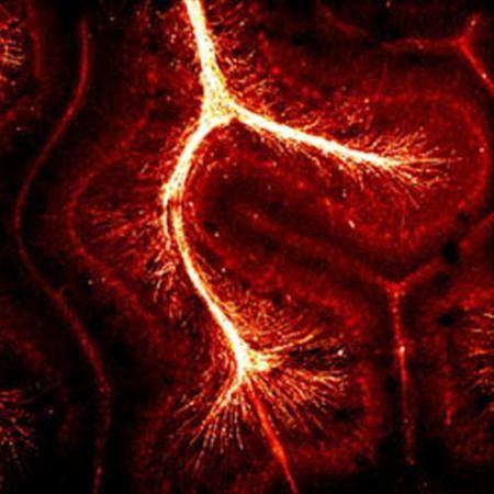
IN PHOTO: An image of superhighways in the brain, with the gold color showing a protein making up myelin, which speeds the conduction of electrical signals along nerve cells, allowing us to think more quickly.
It is interesting to note that infant massage — including Resting Hands and holding methods — increase the myelination of these “superhighways” in the baby’s brain. The “myelin sheath” is like the plastic or rubber coating around an electrical wire. The more the myelin sheath is developed, the less the infant’s over-sensitivity to stimulation.